
Stains
Discoloration on the teeth are referred to as stains.These discolorations vary from yellowish-brown to brownish- black. They vary in shape and size from thin small lines to irregular and blotchy patches.
These stains can be external stains or internal stains
Causes
External stains Internal stains
| 1. Food and drinks | a. Porphyria |
| 2. Nicotine and tobacco products | b. medications |
| 3. Chromogenic bacterial stains | c. erythroblastosis fetalis |
| 4. Tartar | d. Celiac disease |
| 5. Fluorosis | e. Enamel hypoplasia- trauma |
| 6. Metallic components | f. caries |
| 7. Tooth decay | g. nutritional deficiencies |
| h. genetic defects | |
| i. Infections | |
| j. dental restorations | |
| k. Hereditary diseases |
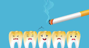
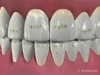

Tobacco stains Enamel hypoplasia white spots on teeth
Grading of stains
| Absent | - |
|---|---|
| Individual spots or continuous line of stains on the gingival margins of tooth on labial and lingual surface of tooth without involvement of proximal surface Or Stains involving only proximal surface Or Individual spots of stains limited to anatomic defects, restoration margins Or orthodontic brackets | + |
| Stains covering up to half of labial or lingual surface of teeth with or without involvement of proximal surface | ++ |
| Stains covering more than half of labial or lingual crown surface, with or without involvement of proximal surface | +++ |
Calculus
Once teeth erupt into the oral cavity various materials gather and accumate on the tooth structures. These accumulations can be hard and soft. The hard tissue accumulates are referred to as calculus
Calculus are mineralized bacterial plaque that forms on the teeth and dental prosthesis
Calculus are further divided into
- Supragingival(on clinical crown or above gingival margin)
- Subgingival ( below gingival margin)

Image showing calculus found above and below the gums
Grading of calculus
| 0 | No calculus |
|---|---|
| + | trace |
| ++ | slight- calculus deposits 1mm or less |
| +++ | Moderate- calculus deposit 1-2mm |
| ++++ | Heavy- calculus deposits more than 2mm |
Gum health

- Gingivitis : inflammation of gums is called gingivitis. Normal colour of the gums is pale pink. In gingivitis, gum turns from red to blue. Bleeds on touch, enlarged and songy in consistency

Image showing reddish swollen gingiva
-
Oral hygiene : depending on the debris and calculus present on the teeth we determine oral hygiene
-
Gum recession: Gum recession- displacement of marginal gums exposing the junction of cementum and enamel
| Class I | Marginal tissue recession that does not extend the mucogingival junction |
| Class II | Marginal tissue recession that extends to or beyond the mucogingival junction, with no periodontal attachment loss ( bone or soft tissue) in the interdental area |
| Class III | Marginal tissue recession that extends to or beyond the mucogingival junction, with periodontal attachment loss in the interdental area or malpositioning of teeth |
| Class IV | Marginal tissue recession that extends to or beyond the mucogingival junction with severe bone or soft-tissue loss in the interdental area and/or severe malpositioning of teeth |

Teeth health
- Tooth decay: it is the destruction of hard and soft tissues of teeth

Tooth stain Enamel caries Dentinal Caries Pulpitis Apical periodontitis
1. Root stumps: root pieces of the tooth present inside the gums

X-ray showing root stumps of lower back teeth
-
Tooth wear : tooth undergoes wear and tear during chewing, night grinding or clenching
-
Attrition: By tooth to tooth grinding or rubbing ( mechanical) the edges of front and back teeth may wear off exposing the inner layers of the teeth

Image showing short lateral incisors
- Abrasion : Scraping off of the tooth structure by forgein particles, this can be seen in single tooth or multiple teeth
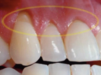
Image showing abrasion of teeth
- Abfraction : Wedge shaped defect in the neck region of tooth

Image showing wedge shaped defect at the neck region of tooth
- Erosion : Loss of dental hard tissue by non bacteriogenic acids

Image showing erosions on upper front teeth
- Missing tooth: loss of teeth can be due to accidents or due to loose gums

Image showing missing upper front teeth
- Non vital tooth: when any tooth is injured during fall or accidents the nerve tissue inside the tooth will be damaged which results in death of this tissue. These type of teeth does not respond to hot or cold and gradually change in color to black
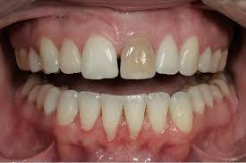
Image showing non vital upper front teeth which is darker in color
Teeth alignment:
- Crowding : overlapping of teeth one above the other. This is can be seen in front teeth as well as back teeth in both upper and lower arches on both the sides

Image showing overlapping of upper front teeth
- Spacing : the gap between the teeth is known as spacing can be seen in front and back teeth

Image showing gap between upper front teeth
- Please mention if any other emergency finding :facial swelling, pus discharge, severe tooth pain or pink tooth
Diagnosis
Nature and cause of the disease
Treatment plan
Procedures or medications given to cure the problems
- Preventive Measures
- Brush your teeth twice daily using tooth brush and fluoridated toothpaste using proper technique
- Clean your tongue using tongue cleaner
- Floss your teeth daily
- Eat healthy and balanced diet
- Reduce sugar intake in between meals
- Avoid carbonated drinks
- Have regular check up with your dentist once in every 6 months
- In case of tooth pain please visit your nearest dentist
- Precautions: In case of dislodged fillings and caps please visit your dentist and ask for replacement failing to do so may cause choking.
- Patient referral